MRI at UBC
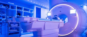
The MRI science community at UBC develops new scientific tools for MRI data acquisition and analysis and shares these new methods collaboratively with scientists at UBC and beyond. Magnetic resonance imaging offers tremendous opportunity and flexibility to develop new, non-invasive techniques that can be performed repeatedly in patients and healthy subjects. The MRI scientists work closely with the UBC MRI Research Centre (Department of Radiology, Faculty of Medicine) which houses two 3 Tesla MRI scanners dedicated to research in human subjects and a preclinical 7 Tesla scanner.

New software release: DECAES for rapid myelin water and luminal water analysis
Alexander Rauscher’s lab is pleased to present DECAES, a free software tool written in Julia. It computes myelin water and luminal water maps in under two minutes. You can find a paper on the method here and the software is freely available on Jon Doucette’s Github page.
News: Mapping cerebrovascular health
We have a gas control system (RespirAct™, Thornhill Medical) that allows us to map cerebrovascular reactivity using our MRI scanners. We can create maps of the brains vascular health that were shown to be useful in a wide range of conditions, such as Alzheimer’s disease, multiple sclerosis, or traumatic brain injury. Please contact Alexander Rauscher’s lab for further details.
MRI Science Faculty
Piotr Kozlowski is a physicist and associate professor in the Department of Radiology. Tumour microenvironment: development and application of MRI based techniques to study tumour blood flow, architecture of the tumour vasculature, tumour tissue oxygen tension, tumour hypoxia, pH, etc. Spinal cord injury: development of an MRI-based technology for very high resolution imaging of rat spinal cord. Current projects include development of specialized RF coils for improved image quality and monitoring changes in myelin in rat spinal cord following injury. Prostate cancer diagnosis: development of a novel MRI-based diagnostic technique for prostate cancer. The technique uses a combination of Diffusion MRI and Contrast Enhanced Dynamic MRI to diagnose and stage prostate cancer. The study involving prostate cancer patients and volunteers is conducted using a research 3T human MRI scanner at the UBC MRI Research Centre.
Shannon Kolind is a physicist and assistant professor in the department of Medicine, division of Neurology. She earned her PhD in Physics at UBC developing myelin water imaging and its application to the study of multiple sclerosis (MS). She then completed a postdoctoral fellowship at the Oxford Centre for Functional MRI of the Brain (FMRIB) at the University of Oxford as well as the Institute of Psychiatry, King’s College London. While in the UK, she specialised in developing new methods to image myelin using MRI, and making them more practical for use in research. She then returned to UBC, this time in the Division of Neurology, to become an Assistant Professor. Shannon’s lab is focused on developing a toolbox of tissue-specific imaging techniques. Her multi-disciplinary team employs these multi-modal tools to achieving greater sensitivity and specificity in clinical research; particularly for clinical trials of new therapies.
Corree Laule is a physicist and assistant professor in the departments of Radiology and Pathology/Lab Medicine. She is interested in understanding the microstructural and pathological determinants which govern T1 and T2 relaxation measures in central nervous system (CNS) tissue. Her primary area of research is multiple sclerosis (MS) and she has extensive experience in imaging both in vivo and post mortem MS brain and spinal cord, with emphasis on characterizing myelin. She also collaborates to study many other CNS applications including schizophrenia, cerebral malaria, bipolar disorder, leukodystrophies and Huntington’s disease, as well the characterization of normal controls. She is particularly interested in myelin and plans to use biochemical analysis and electron microscopy to understand how variations in myelin composition/structure may influence to MRI measures.
Alexander Rauscher is a physicist and associate professor (tenure) and Canada Research Chair in the Department of Pediatrics and also affiliated with the Department of Physics and Astronomy, and the Department of Radiology. His lab’s specific interests are quantitative susceptibility mapping, myelin water imaging, and MR signal formation in the presence of magnetically inhomogeneous tissues, such as nerve fibres or blood vessels. The team is also interested in how the orientation of anisotropic tissue within the MRI scanner’s magnetic field affects the MRI signal. Alex and his group apply their MRI techniques to the investigation of tissue damage and repair in brain injury, for example due to preterm birth, multiple sclerosis, or traumatic brain injury.
Stefan Reinsberg is a physicist and associate professor in the Department of Physics and Astronomy. His group is focused on the development and validation of methods for Cancer Imaging, mostly by MRI. Of particular interest to is the tumour micro-environment and associated tissue heterogeneity. Their experimental techniques include dynamic-contrast-enhanced MRI, diffusion-weighted MRI, dynamic susceptibility-weighted imaging and MR spectroscopy. They use these techniques in clinical (imaging) trials and in pre-clinical research on tumour models. The objective of their work is the identification of imaging biomarkers that can be used clinically for the detection of cancer and monitoring of treatment. An important aspect of this effort is the validation of imaging biomarkers through the use of co-registered histological mapping.
Alexander Weber is an assistant professor in the Department of Pediatrics and an imaging staff scientist at BCCHRI. As a pediatric imaging researcher, his main goals are to better understand MRI contrast mechanisms in order to develop novel imaging and post-processing techniques that aim to improve sensitivity and specificity of biophysical properties of the brain. Furthermore, he aims to use these techniques to better understand differences or changes in white matter in the injured or unhealthy pediatric brain, which can then be used to improve our basic knowledge of the brain, early diagnoses, and to track disease progress with new treatments. Dr. Weber is also interested in creating infrastructure at BCCHRI to provide access and education to clinical researchers who want to engage in using imaging to understand complex neurological and developmental disorders, and to create knowledge translation pathways, such as simple-to-use pipelines in order to better integrate advanced post-processing into the clinical imaging arsenal at BCCH and UBC.
San Xiang is a Professor in the Department of Physics and Astronomy and medical physicist at BC Children’s Hospital specialized in MRI. Besides teaching and servicing, his research activities include technical developments and their clinical applications. Examples are phase-sensitive reconstruction, water-fat imaging, fast imaging with coherent sampling based on signal sparsity, flow quantification and analysis, reduction /correction of artifacts (motion, EPI, banding in bSSFP, metal etc).
Alex MacKay is a physicist and professor emeritus in the Department of Physics and Astronomy. For several decades, he as been working with nuclear magnetic resonance, a powerful technique for investigation of structure and dynamics on an incredibly broad distance scale from the atomic to the macroscopic level. His research has focused on the development of quantitative interpretations of biomedical NMR results in terms of structure, dynamics and composition at the molecular and cellular levels. He and his team have spent considerable effort studying local environments in biological tissue from the water NMR signal. In human brain, they identified a reservoir of water compartmentalized in the myelin component of white matter. These measurements are providing new fundamental insights into the role of myelin in both normal and abnormal brain. Alex MacKay has worked extensively on myelin degeneration in various neurological diseases.